Understanding Flow Cytometry: Key Concepts, Tools, and Uses
Flow cytometry is a powerful analytical technique used extensively in various fields, including immunology, cell biology, and clinical diagnostics. This article aims to provide a comprehensive understanding of flow cytometry, covering its fundamental concepts, essential tools, and diverse applications. By the end of this article, readers will have a solid grasp of how flow cytometry works, its significance in research and clinical settings, and the future directions of this technology.
1. The Fundamentals of Flow Cytometry
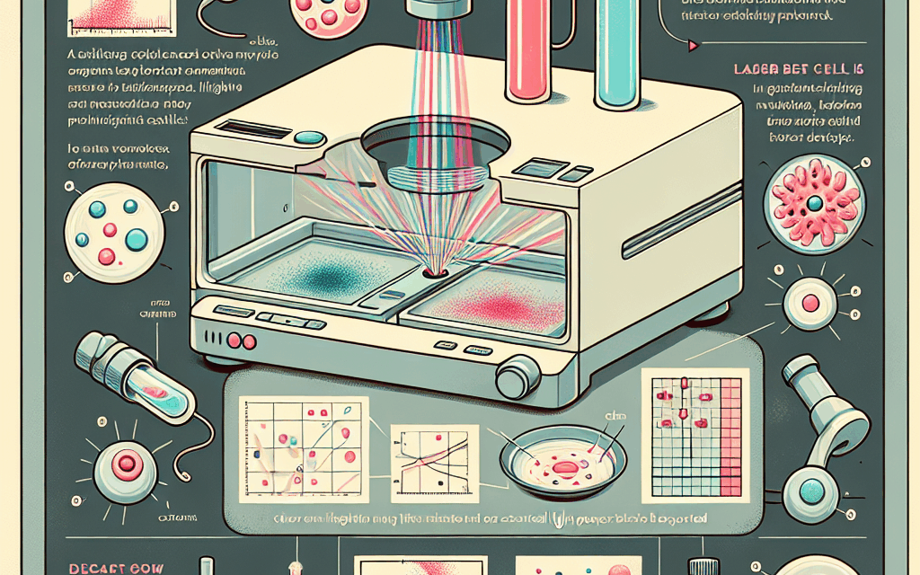
Flow cytometry is a technique that allows for the rapid analysis of physical and chemical characteristics of cells or particles suspended in a fluid stream. The fundamental principle behind flow cytometry is the measurement of light scattering and fluorescence emitted by cells as they pass through a laser beam. This section will delve into the core concepts that underpin flow cytometry, including the principles of light scattering, fluorescence, and the components of a flow cytometer.
1.1 Principles of Light Scattering
Light scattering is a critical phenomenon in flow cytometry. When a laser beam interacts with a particle, such as a cell, it can scatter in different directions. The amount and angle of scattered light provide information about the size and granularity of the cell. There are two primary types of light scattering:
- Forward Scatter (FSC): This measures the light scattered in the forward direction and is primarily related to the size of the cell.
- Side Scatter (SSC): This measures the light scattered at a 90-degree angle and is indicative of the internal complexity or granularity of the cell.
By analyzing FSC and SSC, researchers can differentiate between various cell types based on their size and internal structure.
1.2 Fluorescence in Flow Cytometry
Fluorescence is another key aspect of flow cytometry. Cells can be labeled with fluorescent dyes or antibodies that bind to specific cellular components. When these labeled cells pass through the laser beam, they emit light at specific wavelengths. This emitted light is detected and quantified, allowing for the identification and characterization of different cell populations.
Fluorescent markers can be used to target various cellular components, such as:
- Cell surface markers (e.g., CD4, CD8 in T cells)
- Intracellular proteins (e.g., cytokines)
- Nucleic acids (e.g., DNA, RNA)
The ability to use multiple fluorescent markers simultaneously enables the analysis of multiple parameters in a single experiment, making flow cytometry a highly versatile tool.
1.3 Components of a Flow Cytometer
A flow cytometer consists of several key components that work together to analyze cells:
- Fluidics System: This system transports cells in a fluid stream to the laser beam. It ensures that cells are aligned in a single file for accurate measurement.
- Optics System: This includes lasers and detectors that measure light scattering and fluorescence. Different lasers can excite various fluorescent dyes.
- Electronics System: This converts the detected signals into digital data for analysis and storage.
- Software: Flow cytometry software is used for data analysis, allowing researchers to visualize and interpret the results.
Understanding these components is essential for optimizing flow cytometry experiments and interpreting the data accurately.
2. Tools and Technologies in Flow Cytometry
The evolution of flow cytometry has been marked by significant advancements in technology and instrumentation. This section will explore the various tools and technologies that have enhanced the capabilities of flow cytometry, including the types of flow cytometers, fluorescent dyes, and data analysis software.
2.1 Types of Flow Cytometers
Flow cytometers can be categorized based on their capabilities and applications. The main types include:
- Analytical Flow Cytometers: These are designed for basic analysis of cell populations, typically measuring a limited number of parameters.
- Sorting Flow Cytometers: These advanced systems can separate and collect specific cell populations based on their characteristics, allowing for further analysis or culture.
- High-Throughput Flow Cytometers: These instruments are capable of processing large numbers of samples quickly, making them ideal for large-scale studies.
- Mass Cytometers: Utilizing mass spectrometry, these instruments can analyze hundreds of parameters simultaneously, providing a more comprehensive view of cellular characteristics.
The choice of flow cytometer depends on the specific research question and the complexity of the analysis required.
2.2 Fluorescent Dyes and Antibodies
The selection of appropriate fluorescent dyes and antibodies is crucial for successful flow cytometry experiments. There are several factors to consider:
- Excitation and Emission Wavelengths: Each dye has specific wavelengths for excitation and emission. It is essential to match these with the lasers available on the flow cytometer.
- Brightness and Photostability: Some dyes are brighter and more stable than others, which can affect the sensitivity and reliability of the results.
- Specificity: Antibodies must be validated for specificity to ensure accurate targeting of the intended cellular components.
Commonly used fluorescent dyes include FITC, PE, APC, and their tandem conjugates, each with unique properties suitable for different applications.
2.3 Data Analysis Software
Data analysis is a critical step in flow cytometry, as it transforms raw data into meaningful insights. Several software packages are available for analyzing flow cytometry data, including:
- FlowJo: A widely used software that offers comprehensive tools for data visualization, gating, and statistical analysis.
- FCS Express: This software provides an intuitive interface for data analysis and is known for its flexibility in handling complex datasets.
- R and Bioconductor: For advanced users, R offers packages specifically designed for flow cytometry data analysis, allowing for custom analyses and visualizations.
Choosing the right software depends on the complexity of the data and the specific analysis requirements.
3. Applications of Flow Cytometry in Research
Flow cytometry has a wide range of applications in research, particularly in the fields of immunology, cancer biology, and stem cell research. This section will explore some of the most significant applications of flow cytometry in these areas.
3.1 Immunology
In immunology, flow cytometry is used to analyze immune cell populations, their activation states, and functional responses. Key applications include:
- Characterization of Immune Cell Subsets: Flow cytometry allows researchers to identify and quantify different immune cell types, such as T cells, B cells, and dendritic cells, based on surface markers.
- Assessment of Cytokine Production: By using intracellular staining techniques, researchers can measure the production of cytokines by immune cells, providing insights into their functional status.
- Monitoring Immune Responses: Flow cytometry is instrumental in studying immune responses to infections, vaccines, and therapies, helping to understand how the immune system reacts to various stimuli.
For example, a study published in “Nature” demonstrated how flow cytometry was used to analyze T cell responses in patients receiving immunotherapy for cancer, revealing critical insights into treatment efficacy.
3.2 Cancer Research
Flow cytometry plays a pivotal role in cancer research by enabling the analysis of tumor cells and the immune microenvironment. Key applications include:
- Identification of Cancer Stem Cells: Flow cytometry can be used to isolate and characterize cancer stem cells, which are believed to drive tumor growth and metastasis.
- Assessment of Tumor Heterogeneity: By analyzing multiple markers simultaneously, researchers can study the heterogeneity of tumor cell populations, which is crucial for understanding tumor behavior and treatment resistance.
- Monitoring Minimal Residual Disease (MRD): Flow cytometry is used to detect residual cancer cells after treatment, providing valuable prognostic information.
A notable example is the use of flow cytometry in acute lymphoblastic leukemia (ALL) to monitor MRD, which has been shown to correlate with patient outcomes.
3.3 Stem Cell Research
Flow cytometry is essential in stem cell research, allowing for the identification and characterization of stem cell populations. Key applications include:
- Isolation of Stem Cells: Flow cytometry can be used to sort stem cells based on specific surface markers, enabling researchers to study their properties and potential.
- Assessment of Differentiation: By analyzing changes in marker expression, researchers can track the differentiation of stem cells into specialized cell types.
- Functional Assays: Flow cytometry can be used to assess the functional capabilities of stem cells, such as their ability to proliferate or respond to stimuli.
For instance, a study published in “Cell Stem Cell” utilized flow cytometry to analyze the differentiation of induced pluripotent stem cells (iPSCs) into cardiomyocytes, providing insights into cardiac development.
4. Clinical Applications of Flow Cytometry
Flow cytometry is not only a research tool but also has significant clinical applications, particularly in diagnostics and monitoring of diseases. This section will explore how flow cytometry is used in clinical settings, including hematology, oncology, and infectious diseases.
4.1 Hematology
In hematology, flow cytometry is widely used for the diagnosis and monitoring of blood disorders. Key applications include:
- Diagnosis of Leukemias and Lymphomas: Flow cytometry is essential for identifying specific cell populations in blood and bone marrow, aiding in the diagnosis of various hematological malignancies.
- Assessment of Hemoglobin Variants: Flow cytometry can be used to analyze different hemoglobin types, which is crucial for diagnosing conditions like sickle cell disease and thalassemia.
- Monitoring Treatment Response: By assessing changes in cell populations, flow cytometry can help monitor the effectiveness of treatments for blood disorders.
A study published in “Blood” highlighted the role of flow cytometry in diagnosing acute myeloid leukemia (AML) by identifying specific immunophenotypes associated with the disease.
4.2 Oncology
Flow cytometry is increasingly used in oncology for various applications, including:
- Biomarker Discovery: Flow cytometry can identify novel biomarkers associated with cancer progression, aiding in the development of targeted therapies.
- Assessment of Tumor Microenvironment: By analyzing immune cell populations within tumors, researchers can gain insights into the tumor microenvironment and its impact on treatment response.
- Clinical Trials: Flow cytometry is often used in clinical trials to monitor patient responses to new therapies, providing valuable data for evaluating treatment efficacy.
For example, a clinical trial published in “The Lancet” utilized flow cytometry to assess immune responses in patients receiving checkpoint inhibitors for melanoma, demonstrating its utility in evaluating therapeutic outcomes.
4.3 Infectious Diseases
Flow cytometry is also employed in the diagnosis and monitoring of infectious diseases. Key applications include:
- Detection of Pathogen-Specific Immune Responses: Flow cytometry can measure immune responses to specific pathogens, providing insights into host-pathogen interactions.
- Monitoring HIV Infection: Flow cytometry is used to quantify CD4+ T cell counts in HIV-infected individuals, which is crucial for monitoring disease progression and treatment efficacy.
- Vaccine Development: Flow cytometry plays a role in evaluating immune responses to vaccines, helping to assess their effectiveness.
A study published in “Nature Medicine” demonstrated how flow cytometry was used to analyze immune responses in individuals vaccinated against COVID-19, providing critical data on vaccine efficacy.
5. Future Directions in Flow Cytometry
The field of flow cytometry is continuously evolving, with advancements in technology and methodologies paving the way for new applications and improved capabilities. This section will explore some of the future directions in flow cytometry, including innovations in instrumentation, data analysis, and emerging applications.
5.1 Innovations in Instrumentation
Future advancements in flow cytometry instrumentation are expected to enhance its capabilities significantly. Key innovations include:
- Increased Multiplexing: New technologies are being developed to allow for the simultaneous analysis of more parameters, enabling a more comprehensive understanding of cellular characteristics.
- Miniaturization: Advances in microfluidics are leading to the development of smaller, portable flow cytometers that can be used in field settings or resource-limited environments.
- Integration with Other Technologies: Combining flow cytometry with other techniques, such as mass spectrometry or imaging, can provide complementary data and enhance the overall analysis.
These innovations will expand the applications of flow cytometry and make it more accessible to researchers and clinicians.
5.2 Advances in Data Analysis
The increasing complexity of flow cytometry data necessitates advancements in data analysis techniques. Future directions include:
- Machine Learning and AI: The integration of machine learning algorithms can improve data analysis by automating gating strategies and identifying patterns in complex datasets.
- Cloud-Based Analysis: Cloud computing can facilitate data sharing and collaborative analysis, allowing researchers to access and analyze large datasets more efficiently.
- Standardization of Data Reporting: Efforts are underway to standardize data reporting formats, making it easier to compare results across studies and laboratories.
These advancements will enhance the reproducibility and reliability of flow cytometry data, ultimately benefiting research and clinical applications.
5.3 Emerging Applications
As flow cytometry technology continues to evolve, new applications are emerging across various fields. Some notable areas include:
- Single-Cell Genomics: Flow cytometry is being integrated with genomic techniques to analyze gene expression at the single-cell level, providing insights into cellular heterogeneity.
- Microbiome Analysis: Flow cytometry can be used to study microbial communities, helping to understand their roles in health and disease.
- Personalized Medicine: Flow cytometry is playing a role in the development of personalized therapies by analyzing patient-specific cellular responses to treatments.
These emerging applications highlight the versatility of flow cytometry and its potential to address complex biological questions.
Conclusion
Flow cytometry is a powerful and versatile tool that has transformed the fields of research and clinical diagnostics. By understanding its fundamental principles, tools, and diverse applications, researchers and clinicians can harness its capabilities to gain valuable insights into cellular characteristics and behaviors. As technology continues to advance, flow cytometry will undoubtedly play an increasingly important role in scientific discovery and patient care.
In summary, the key takeaways from this article include:
- The fundamental principles of flow cytometry, including light scattering and fluorescence.
- The various types of flow cytometers and their applications in research and clinical settings.
- The significant role of flow cytometry in immunology, cancer research, and stem cell biology.
- The clinical applications of flow cytometry in hematology, oncology, and infectious diseases.
- The future directions of flow cytometry, including innovations in instrumentation, advances in data analysis, and emerging applications.
As we look to the future, flow cytometry will continue to evolve, providing researchers and clinicians with powerful tools to explore the complexities of biology and improve patient outcomes.


