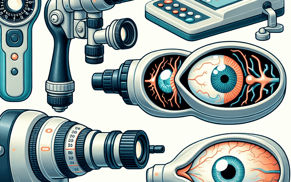
5 Diagnostic Tools to Identify the Causes of Eye Bulging
Eye bulging, medically known as exophthalmos or proptosis, is a condition characterized by the protrusion of one or both eyes. This condition can be alarming for patients and may indicate underlying health issues, ranging from thyroid disorders to tumors. Accurate diagnosis is crucial for effective treatment. In this article, we will explore five diagnostic tools that healthcare professionals use to identify the causes of eye bulging, providing insights into their methodologies, applications, and significance.
1. Comprehensive Eye Examination
A comprehensive eye examination is often the first step in diagnosing the cause of eye bulging. This examination typically includes a series of tests designed to assess the overall health of the eyes and surrounding structures.
Visual Acuity Tests
Visual acuity tests measure how well a person can see at various distances. These tests help determine if the bulging is affecting vision. The Snellen chart is commonly used, where patients read letters from a distance. A significant decrease in visual acuity may indicate pressure on the optic nerve, often seen in conditions like Graves’ disease.
Ocular Motility Assessment
Ocular motility tests evaluate the movement of the eyes. The healthcare provider will ask the patient to follow a moving object in various directions. Limited eye movement can suggest muscle involvement, which is common in thyroid eye disease. For instance, in Graves’ disease, inflammation can affect the extraocular muscles, leading to restricted movement and bulging.
Intraocular Pressure Measurement
Measuring intraocular pressure (IOP) is crucial, as elevated IOP can indicate glaucoma, which may coexist with other conditions causing eye bulging. Tonometry is the standard method for measuring IOP, and abnormal readings can prompt further investigation into the underlying causes.
Fundoscopy
Fundoscopy involves examining the interior of the eye, particularly the retina and optic nerve. This examination can reveal signs of increased intracranial pressure or other abnormalities that may contribute to eye bulging. For example, swelling of the optic disc can indicate papilledema, often associated with conditions like tumors or severe hypertension.
Case Study: Thyroid Eye Disease
A 45-year-old female patient presented with bilateral eye bulging and visual disturbances. A comprehensive eye examination revealed reduced visual acuity and restricted ocular motility. Fundoscopy showed signs of optic nerve swelling. Further tests confirmed a diagnosis of Graves’ disease, leading to appropriate treatment.
2. Imaging Studies
Imaging studies play a vital role in diagnosing the causes of eye bulging. They provide detailed views of the structures behind the eyes, helping to identify tumors, inflammation, or other abnormalities.
Computed Tomography (CT) Scans
CT scans are particularly useful for visualizing the orbits and surrounding tissues. They can help identify masses, such as tumors or cysts, and assess the extent of any inflammation. For instance, a CT scan may reveal an enlarged thyroid gland or retro-orbital fat, common in thyroid eye disease.
Magnetic Resonance Imaging (MRI)
MRI is another powerful imaging tool that provides detailed images of soft tissues. It is especially useful for evaluating the optic nerve and surrounding structures. MRI can help differentiate between various causes of eye bulging, such as tumors versus inflammatory conditions. For example, an MRI may show a mass compressing the optic nerve, indicating a need for surgical intervention.
Ultrasound Imaging
Ultrasound is a non-invasive imaging technique that can be used to assess the orbits. It is particularly useful for evaluating the size and structure of the eye and surrounding tissues. Ultrasound can help identify fluid collections or masses that may be causing bulging. For instance, a patient with a retro-orbital hematoma may show significant bulging on ultrasound.
Case Study: Orbital Tumor
A 60-year-old male patient presented with unilateral eye bulging. A CT scan revealed a well-defined mass in the orbit, consistent with a meningioma. An MRI confirmed the diagnosis, leading to surgical intervention. The imaging studies were crucial in guiding the treatment plan.
3. Blood Tests
Blood tests are essential for diagnosing systemic conditions that may lead to eye bulging. They can help identify hormonal imbalances, autoimmune disorders, and other underlying health issues.
Thyroid Function Tests
Thyroid function tests measure levels of thyroid hormones (T3, T4) and thyroid-stimulating hormone (TSH). Abnormal levels can indicate hyperthyroidism or hypothyroidism, both of which can lead to eye bulging. For example, elevated T3 and T4 levels with suppressed TSH are indicative of Graves’ disease, a common cause of exophthalmos.
Autoimmune Markers
In cases where autoimmune disorders are suspected, specific blood tests can help identify markers associated with conditions like Graves’ disease or Hashimoto’s thyroiditis. These tests may include thyroid receptor antibodies (TRAb) and thyroglobulin antibodies (TgAb). Positive results can confirm an autoimmune etiology for the eye bulging.
Complete Blood Count (CBC)
A complete blood count can help identify signs of infection or inflammation. Elevated white blood cell counts may suggest an infectious process, while anemia may indicate chronic disease. For instance, a patient with orbital cellulitis may present with elevated white blood cell counts and eye bulging due to inflammation.
Case Study: Autoimmune Thyroid Disease
A 35-year-old female patient with a history of hyperthyroidism presented with bilateral eye bulging. Blood tests revealed elevated T3 and T4 levels, along with positive TRAb. The diagnosis of Graves’ disease was confirmed, leading to treatment with antithyroid medications.
4. Biopsy and Cytology
In some cases, a biopsy may be necessary to determine the cause of eye bulging, especially when a mass is suspected. Biopsy and cytology provide definitive diagnoses by allowing for histological examination of tissue samples.
Fine Needle Aspiration Biopsy (FNAB)
FNAB is a minimally invasive procedure used to obtain tissue samples from masses in the orbit. It is particularly useful for diagnosing tumors or inflammatory lesions. The procedure involves using a thin needle to extract cells from the suspicious area, which are then examined under a microscope. For example, FNAB can help differentiate between benign and malignant tumors in the orbit.
Excisional Biopsy
In cases where FNAB is inconclusive, an excisional biopsy may be performed. This involves surgically removing the entire mass for examination. Excisional biopsies are often necessary for larger tumors or when there is a high suspicion of malignancy. For instance, a patient with a suspected orbital lymphoma may undergo an excisional biopsy to confirm the diagnosis.
Case Study: Orbital Lymphoma
A 50-year-old male patient presented with unilateral eye bulging and a palpable mass in the orbit. FNAB was performed, revealing atypical lymphoid cells. An excisional biopsy confirmed the diagnosis of orbital lymphoma, leading to appropriate treatment with chemotherapy.
5. Endocrine Evaluation
Endocrine evaluation is crucial for diagnosing conditions related to hormonal imbalances that may cause eye bulging. This evaluation often involves a multidisciplinary approach, including endocrinologists and ophthalmologists.
Thyroid Imaging and Function Tests
In addition to blood tests, imaging studies of the thyroid gland may be performed to assess its size and structure. Thyroid scans using radioactive iodine can help determine if the gland is overactive, which is common in Graves’ disease. This evaluation is essential for understanding the relationship between thyroid function and eye bulging.
Assessment of Other Endocrine Disorders
Other endocrine disorders, such as Cushing’s syndrome or acromegaly, can also lead to eye bulging. Evaluating hormone levels related to these conditions is crucial. For instance, elevated cortisol levels in Cushing’s syndrome can lead to fat deposition around the eyes, causing bulging.
Case Study: Cushing’s Syndrome
A 40-year-old female patient presented with bilateral eye bulging and weight gain. Endocrine evaluation revealed elevated cortisol levels and a diagnosis of Cushing’s syndrome. Treatment focused on addressing the underlying cause, leading to resolution of the eye bulging.
Conclusion
Eye bulging can be a distressing symptom with various underlying causes. Accurate diagnosis is essential for effective treatment and management. The five diagnostic tools discussed in this article—comprehensive eye examinations, imaging studies, blood tests, biopsy and cytology, and endocrine evaluations—play a crucial role in identifying the causes of eye bulging.
Healthcare professionals must utilize a combination of these tools to arrive at a definitive diagnosis. Early detection and intervention can significantly improve patient outcomes, particularly in cases of serious underlying conditions such as tumors or autoimmune disorders. By understanding the diagnostic process, patients can be better informed about their health and the steps necessary for effective treatment.
In summary, if you or someone you know is experiencing eye bulging, it is essential to seek medical attention promptly. A thorough evaluation using these diagnostic tools can lead to a better understanding of the condition and appropriate management strategies.


